The 2026 Nobel Prize in Physiology or Medicine was jointly awarded to three scientists who created optogenetics, a technique for activating and silencing nerve cells with light. On October 5, the Nobel Assembly at Sweden’s Karolinska Institutet named Karl Deisseroth, Peter Hegemann, and Georg Nagel as joint laureates.
The prize was awarded for discoveries concerning light-gated ion channels and optogenetics. The research began with a protein that makes green algae swim toward light.
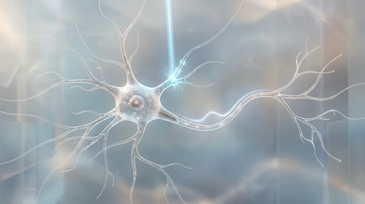 Concept image of a nerve cell firing a signal under blue light — Source: concept image · self-generated with agy
The laureates and the prize
| Laureate | Affiliation | Born |
|---|---|---|
| Karl Deisseroth | HHMI, Stanford University (US) | 1971 |
| Peter Hegemann | Humboldt University of Berlin (Germany) | 1954 |
| Georg Nagel | University of Würzburg (Germany) | 1953 |
The prize money of about ₩1.7 billion (12 million Swedish kronor, about $1.2 million) is split equally among the three. The Nobel Assembly said Hegemann did the prize-winning work while at the Max Planck Institute of Biochemistry and Nagel while at the Max Planck Institute of Biophysics.
Per Svenningsson, chair of the Nobel Committee, said optogenetics provides opportunities to analyze brain function in ways that were previously impossible.
From discovery to the prize
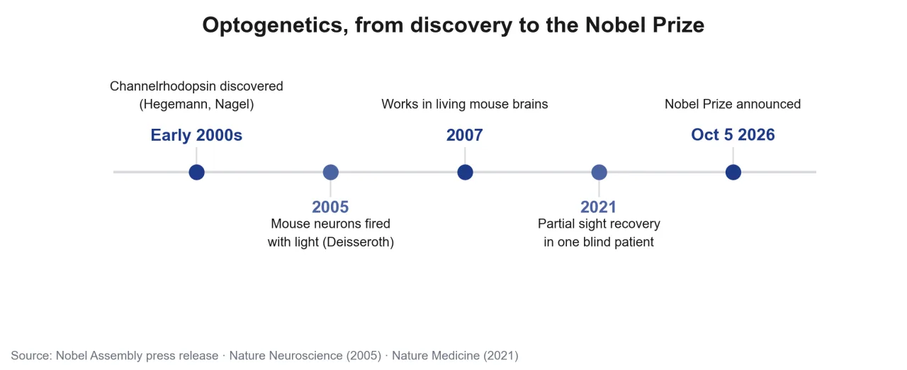 Key years from the discovery of optogenetics to the Nobel Prize — Source: self-rendered from the Nobel Assembly press release and papers
It started with the single-celled green alga Chlamydomonas. Hegemann wondered how this alga swims toward light, and in the early 2000s he and Nagel discovered a protein called channelrhodopsin on the cell surface. When it receives blue light, a channel opens in the protein and charged ions flow into the cell, producing an electrical signal.
The two also confirmed that introducing this protein into any cell makes that cell respond to light. According to the Nobel Assembly press release, in 2005 Deisseroth succeeded in introducing the channelrhodopsin gene into mouse nerve cells and triggering nerve signals with blue light, and two years later made it work in the brains of living mice.
The 2005 paper
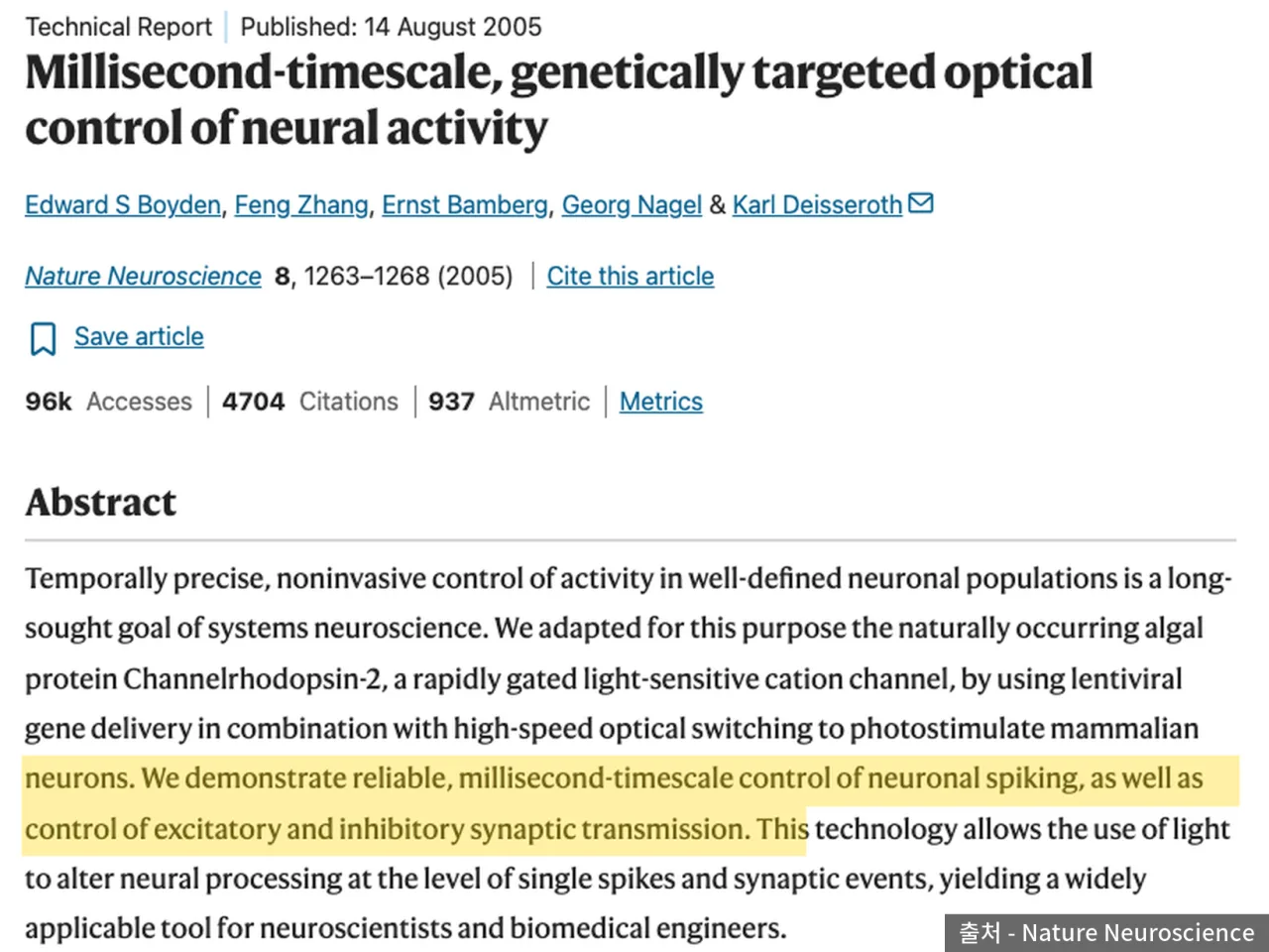 Title and abstract of the 2005 Nature Neuroscience paper — Source: Nature Neuroscience
That result is Millisecond-timescale, genetically targeted optical control of neural activity, published in Nature Neuroscience in August 2005. Edward Boyden was first author, Deisseroth was corresponding author, and laureate Nagel was also a co-author.
The researchers wrote in the abstract that they introduced the channelrhodopsin-2 gene into mammalian nerve cells with a lentivirus and, by switching light on and off rapidly, controlled the firing of nerve cells on a millisecond (one-thousandth of a second) scale. They could even control excitatory and inhibitory synaptic transmission with light. According to the Nature page, the paper has been cited 4,704 times.
How optogenetics works
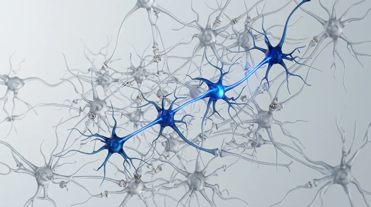 Concept image of a brain circuit in which only specific nerve cells respond to light — Source: concept image · self-generated with agy
[Gene delivery] Introduce the gene for a light-sensing protein only into a specific type of nerve cell
[Light stimulation] Shine light on that area to activate or silence only those nerve cells
Earlier methods inferred function by damaging or electrically stimulating a particular brain area, but neighboring cells were affected too, making it hard to prove cause and effect. Optogenetics can activate or silence only a specific type of cell at a chosen moment, so experiments can confirm whether those cells are directly involved in memory, emotion, or behavior.
Human treatment cases
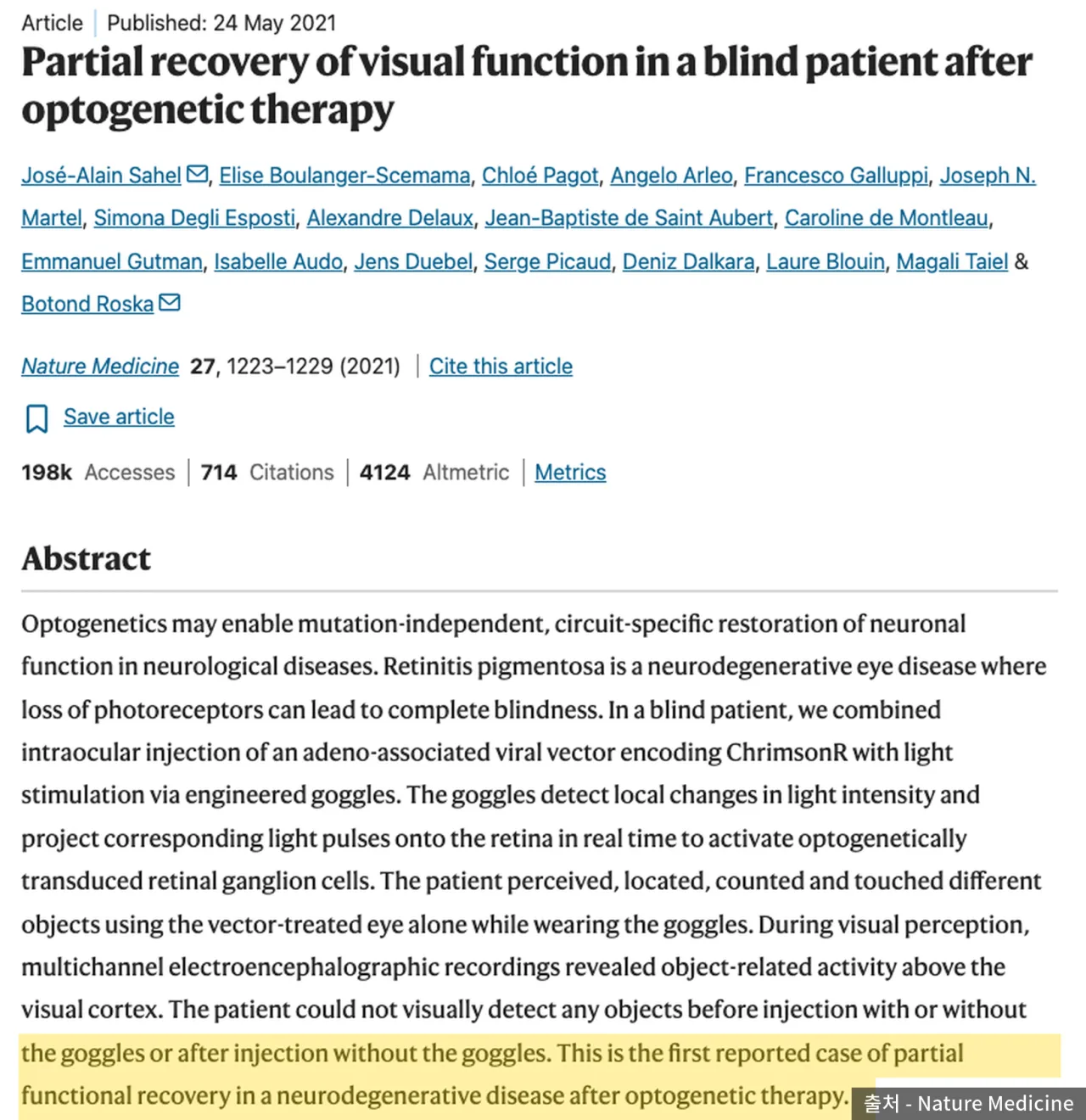 Abstract of the 2021 Nature Medicine paper on partial recovery of visual function — Source: Nature Medicine
The Nobel Assembly noted that in clinical medicine, optogenetics is being used in attempts to restore sight to blind people. A leading example is the paper by José-Alain Sahel and Botond Roska’s team published in Nature Medicine in May 2021.
They injected a viral vector carrying the gene for the light-sensing protein ChrimsonR into one eye of a single patient who had lost sight to retinitis pigmentosa, and fitted goggles that detect changes in ambient brightness and project light signals onto the retina. Wearing the goggles, the patient could recognize, locate, count, and touch objects using that eye alone, and brain activity responding to objects was recorded over the visual cortex. The authors wrote that it was the first reported case of partial functional recovery after optogenetic therapy in a neurodegenerative disease.
Why it isn’t a treatment yet
Alongside news of the prize, it’s easy to read optogenetics as about to become a treatment for brain diseases, but human application is at an early stage.
| Constraint | Details |
|---|---|
| Gene delivery | Requires inserting genes into human cells with a viral vector |
| Light delivery | Deep brain areas require implanting devices such as optical fibers |
| Clinical evidence | The 2021 sight-recovery report is a single patient |
| Level of recovery | Object recognition with goggles, not normal vision |
The eye became the first clinical target because light can enter it from outside. Brain diseases require placing a light-delivery device inside the head, so more validation is needed before it moves from research tool to treatment. The Nobel Assembly, too, based the prize on developing a way to reveal how the brain works, not on treatment outcomes.
Research in Korea
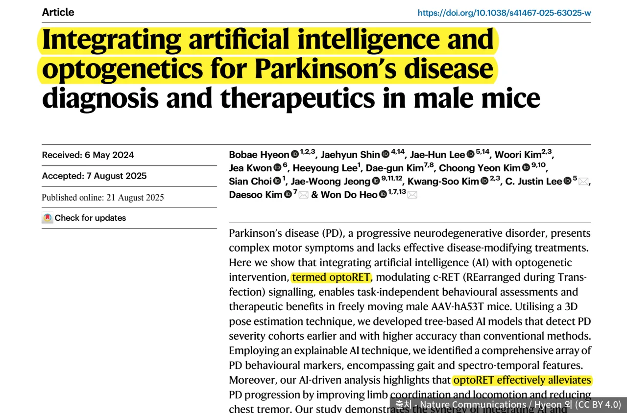 Title and abstract of the 2025 Nature Communications paper by the KAIST–IBS team — Source: Nature Communications / Hyeon et al. (CC BY 4.0)
In Korea, Professor Heo Won-do’s team in the Department of Biological Sciences at KAIST has steadily developed optogenetic tools that control protein activity with light. In August 2025, together with Professor Kim Dae-soo of KAIST and Director Lee C. Justin’s team at the Institute for Basic Science (IBS), they published Parkinson’s disease research in Nature Communications.
Combining optoRET, a technique that activates a neuroprotective signal (c-RET) with light, with AI 3D pose estimation, they distinguished severity in Parkinson’s model mice earlier than existing tests using behavioral indicators such as gait, limb coordination, and tremor, and confirmed that symptom progression was eased. However, the subjects were male mice, not humans.
Sources
- [The Nobel Prize] Press release: The Nobel Prize in Physiology or Medicine 2026 (2026-10-05, laureates, affiliations, achievements, prize money)
- [Nature Neuroscience] Boyden, Zhang, Bamberg, Nagel, Deisseroth, Millisecond-timescale, genetically targeted optical control of neural activity, 8, 1263–1268 (2005; source of 1 image in the body)
- [Nature Medicine] Sahel et al., Partial recovery of visual function in a blind patient after optogenetic therapy, 27, 1223–1229 (2021; source of 1 image in the body)
- [Nature Communications] Hyeon et al. (KAIST, IBS), Integrating artificial intelligence and optogenetics for Parkinson’s disease diagnosis and therapeutics in male mice (2025-08-21, CC BY 4.0; source of 1 image in the body)
- [Scientific American] 2026 Nobel Prize coverage (prize money in dollars)
- Exchange rate: converted at $1 ≈ ₩1,400